Professor Washington University/B-JH/SLCH Consortium PM&R Program Saint Louis, Missouri
Disclosure(s):
Gloria Coden, MD: No financial relationships to disclose
Case Diagnosis: 34-year-old female presented with 19 months of bilateral lower extremity weakness after giving birth and found to have type 1 myotonic dystrophy on electromyography.
Case Description or Program Description: 34-year-old female with a past surgical history of a T3-L4 posterior spinal fusion 13 years ago who presented with 19 months of bilateral lower extremity weakness after giving birth. Patient reports difficulty running and jumping. Patient denies any pain. Physical exam demonstrated 5/5 strength in bilateral upper extremities, dorsiflexion and toe extension, 4/5 bilateral knee extension, 3+/5 bilateral hip flexion and plantarflexion. Radiographs demonstrated intact hardware without loosening. Magnetic resonance imaging demonstrated mild facet degenerative changes without neuroforaminal or spinal stenosis. Patient was referred for nerve conduction studies (NCS) and electromyography due to concern for spine pathology.
Setting: Outpatient physical medicine and rehabilitation office at an academic medical center
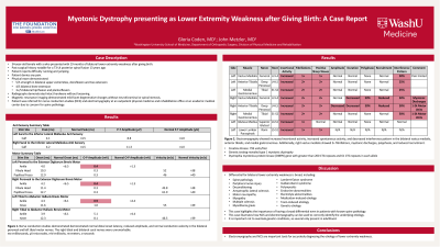
Assessment/Results: NCS demonstrated normal distal onset latency, reduced amplitude, and normal conduction velocity in the bilateral peroneal and left tibial motor nerves. The right tibial and bilateral sural nerves were unremarkable. Electromyography showed increased insertional activity, increased spontaneous activity, and decreased interference pattern in the bilateral vastus medialis, anterior tibialis, and medial gastrocnemius. Additionally, right vastus medialis showed 2+ fibrillations, myotonic discharges, polyphasia, and reduced recruitment. Genetic testing revealed type 1 myotonic dystrophy with greater than 200 repeats and 61 repeats in the DMPK gene.
Discussion (relevance): Differential for bilateral lower extremity weakness is broad, including spine pathology, peripheral nerve injury, deconditioning, amyotrophic lateral sclerosis, motor neuropathy, myopathy, multiple sclerosis, myasthenia gravis, Lambert-Eaton syndrome, Guillain-Barré syndrome, polymyositis, endocrine abnormalities, electrolyte abnormalities, medication-induced, toxin-induced, or genetic etiology. This case illustrates how NCS and electromyography can be used to correctly identify the underlying etiology. It is important not to overlook genetic conditions, as several only present in adulthood.
Conclusions: Electromyography and NCS are important tools for accurately diagnosing the etiology of lower extremity weakness.

.jpg)