Assistant Professor Touro university Newport Beach, California
Disclosure(s):
Aiden Le Roux: No financial relationships to disclose
Background and/or Objectives: Occipital Neuralgia is a relatively rare, but debilitating headache condition that affects ~3 people per 100,000 people in the United States. It is well known that it can be treated with nerve blocks; however, little research exists comparing standardized treatment methods to optimize patient outcomes. Our study looks to provide a replicable, safe, and effective method for using ultrasound to find the greater occipital nerve for occipital neuralgia nerve blocks.
Design: We designed a standardized technique to visualize the occipital nerve with ultrasound, and used it on 3 patients. We confirmed our anatomy with the occipital artery which we were able to palpate.
Setting : Outpatient
Participants : 3 patients
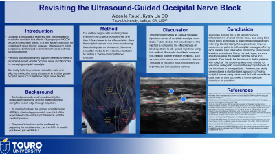
Interventions: Our method begins with localizing 3mm inferior to the occipital protuberance, and then 1.5mm lateral to the affected side. Once the occipital vessels have been found using the color Doppler on ultrasound, the nerve should be medial to the vessels, visualized by finding a “honey-comb” patterned structure. Using this technique, we were able to visualize the greater occipital nerve in 3 patients.
Main Outcome Measures: Visualization of the occipital nerve.
Results: Our study proposes a replicable method for ultrasound-guided GON blocks. The nerve is located 3 mm inferior to the occipital protuberance and 1.5 mm lateral to the affected side. Using color Doppler, the “honeycomb” nerve lies medially to the occipital vessels.
Conclusions: This standardized approach enhances reproducibility and clinical effectiveness. Ultrasound improves precision, reduces complications, and aids diagnosis. By targeting inflamed nerves, it offers a more reliable and effective alternative to blind techniques. Standardization could optimize outcomes, providing reliable pain relief while minimizing invasive procedures.

.jpg)
.jpeg.jpg "Aiden Le Roux (he/him/his) photo")