George Raum, DO: No financial relationships to disclose
Background and/or Objectives: Altered loads at the patellofemoral joint (PFJ) are thought to be the primary contributor to patellofemoral pain syndrome (PFPS) but anatomical correlates are poorly understood. Shear wave elastography (SWE), a technique that quantifies tissue elasticity, may detect viscoelastic changes in key structures. This study aimed to determine the reliability and stability of B-mode and SWE US measures of PFJ structures, and to assess differences present in PFPS patients.
Design: Prospective, case-control study.
Setting : Sports Medicine Clinic
Participants : 19 PFPS patients and 13 controls.
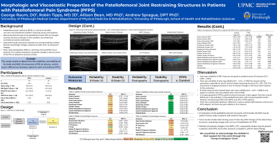
Interventions: Participants underwent US imaging of the vastus lateralis (VL), rectus femoris (RF), vastus intermedius (VI), vastus medialis (VM), quadriceps (QT) and patellar tendon (PT), medial (MFPL) and lateral patellofemoral ligaments (LPFL) and Iliotibial band (ITB). A subset returned within 48 hours to assess stability.
Main Outcome Measures: Reliability and stability of B-mode and SWE imaging assessed via intra-class correlation coefficients, between-group comparisons of tissue measurements assessed via t-tests and effect size.
Results: B-mode inter-rater reliability was poor (ICC=0.27-0.49) for all structures, except for PT thickness and cross-sectional area (CSA) (ICC=0.51-0.69), which had moderate agreement. SWE reliability was excellent across all structures (ICC=0.90-0.991). B-mode stability was poor for PT and ITB thickness (ICC=0.21-0.41), moderate for QT CSA and QT, MPFL, and LPFP thickness and (ICC=0.51-0.71), and good for PT CSA (ICC=0.82). SWE stability was poor for the VL, QT, LPFL and ITB (ICC=-0.50-0.50) and moderate for the RF, VI, VM, PT and MPFL (ICC=0.66-0.75). There were no significant differences between the controls and the most symptomatic limb of the PFPS group, but moderate effect sizes were observed for PT thickness and MPFL and LPFL shear wave speed.
Conclusions: SWE demonstrates excellent reliability, with variable stability while B-mode imaging demonstrated the opposite pattern. Differences in tissue elasticity, especially in the MPFL and PT, suggest SWE may detect meaningful viscoelastic changes relevant to PFPS and warrant further investigation.

.jpg)